The inner workings of the brain’s “sewage system” are finally coming into focus after years of speculation.
For the first time, scientists have visualized the complex plumbing network within the folds of five human brains—a system that had previously only been observed in mice and hypothesized in humans.
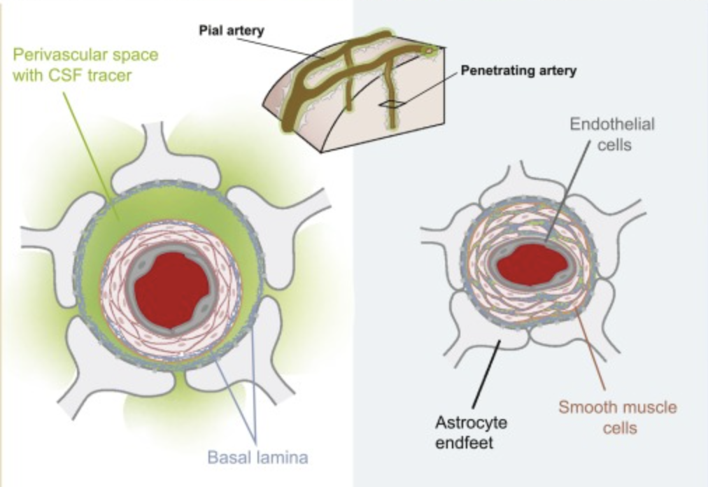
These findings confirm the existence of the glymphatic system, first discovered in mice brains in 2012. This system helps transport cerebrospinal fluid (CSF), which surrounds the brain, into its interior, delivering nutrients and clearing out waste products, including proteins linked to Alzheimer’s disease.
Since its discovery in mice, studies have suggested the glymphatic system also exists in humans, but direct visualization of CSF movement from the brain’s surface into the spaces between neurons has been debated.
“I was skeptical myself, and many still are,” says neurologist Juan Piantino from OHSU. “That’s what makes this discovery so remarkable.”
Piantino and his team at OHSU are the first to capture images of the clear liquid moving through a living human brain, confirming earlier studies that had only offered brief glimpses.
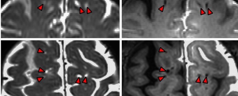
This breakthrough was made possible through the involvement of five adults undergoing brain surgery, which required the diversion of CSF. Before replenishing the fluid, researchers introduced a dark contrast tracer. Using specialized MRI techniques, they tracked the fluid’s movement through the participants’ brains.
The results showed that the brain doesn’t absorb CSF randomly; the fluid follows blood vessels deep into neural tissue. CSF flows through defined channels that wrap around blood vessels, with brain cells forming permeable barriers using their ‘endfeet.’
Some researchers propose that this membrane allows CSF to mix with the protective fluid around brain cells, supplying nutrients and removing waste.
OHSU neurosurgeon Erin Yamamoto explains that in the MRI images, “you can actually see dark perivascular spaces in the brain turn bright,” showing the tracer’s movement, similar to what had been seen in mice.
Although these CSF pathways had been observed in human brains before, OHSU’s MRI scans—taken 12, 24, and 48 hours post-surgery—offered unprecedented insight into the fluid dynamics within the brain.
The study demonstrates that CSF channels are not static but function as “dynamic conduits” that help distribute fluid throughout the brain.
“People suspected these perivascular spaces were important, but it had never been proven,” says Piantino. “Now it has.”
The research was published in PNAS.